


Understanding Fine-Needle Aspiration Cytology (FNAC),
Fine-Needle Aspiration Cytology (FNAC), often simply referred to as Fine-Needle Aspiration (FNA), is a widely used, minimally invasive diagnostic procedure. It involves using a very thin, hollow needle and a syringe to extract a sample of cells, tissue, or fluid from a suspicious mass, lump, or abnormal area within the body. This sample is then prepared on a slide and sent to a cytology laboratory for microscopic examination. FNAC is generally quick, relatively inexpensive, and offers high accuracy when performed and analyzed appropriately. It plays a critical role in modern medicine, particularly in the rapid diagnosis and management of various conditions, including cancer and inflammatory diseases.
Purpose and Clinical Applications
The primary purpose of the FNAC procedure is to determine the nature of a newly identified mass or lesion - specifically, whether it is benign (non-cancerous), malignant (cancerous), or due to an inflammatory process. It is a vital tool for the initial investigation of lumps in readily accessible areas of the body, and increasingly, in deeper lesions with the aid of imaging guidance.
FNAC is most commonly used to biopsy masses in the:
Thyroid: Assessing thyroid nodules to determine if they require further intervention.
Breast: Investigating breast cysts or newly identified solid lumps.
Lymph Nodes: Evaluating enlarged lymph nodes to diagnose infection, inflammation, or metastatic cancer spread.
Salivary Glands
Skin and soft tissues
With advancements in imaging technology, FNAC can now be guided by techniques such as ultrasound, Computed Tomography (CT) scans, or endoscopic ultrasound (EUS) to safely obtain samples from deeper internal organs, including the pancreas, liver (for focal lesions or suspected metastases), and gastrointestinal tract.
Beyond initial diagnosis, FNAC is also employed in the management of known tumors to assess the effect of treatment or to obtain tissue for special molecular and genetic studies that can guide personalized cancer therapies. Furthermore, the technique can be used therapeutically. For instance, therapeutic fine-needle aspiration can drain fluid from abscesses (buildup of pus), cysts (fluid-filled sacs), and seromas (fluid collections common after surgery), often providing a less invasive alternative to surgical incision and drainage, especially in cosmetically sensitive areas like the breast.
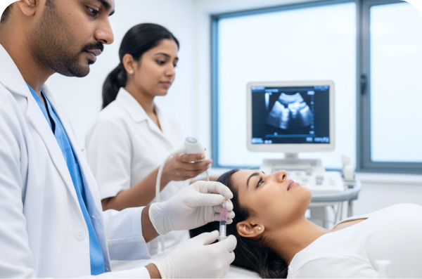
The Procedure: What to Expect
FNAC is an outpatient procedure, typically performed in a clinic, office, or hospital setting. It generally takes about 15 to 30 minutes from start to finish, although the actual sampling process is very brief.
Preparation: In most cases, no special preparation or fasting is required. However, patients should inform their healthcare provider about any blood-thinning medications they are taking, as these might need temporary adjustment before the procedure.
Positioning and Sanitization: The patient is positioned comfortably (lying down or sitting), and the area of the body where the lump is located is thoroughly cleaned with an antiseptic solution.
Anesthesia (Optional): Local anesthesia is sometimes used, especially for deeper or more sensitive areas, but often the discomfort is so minor that it is not required.
Sampling: The healthcare provider, often a cytopathologist or a radiologist, inserts a very thin (narrow gauge, typically 25-22G) needle attached to a syringe into the targeted mass. If the mass is deep or difficult to palpate, imaging guidance (like ultrasound) is used to ensure accurate placement of the needle tip. The syringe is used to create suction (aspiration) to pull out cells, tissue fragments, and fluid. Multiple samples (passes) may be taken from different parts of the lesion to ensure adequate representation of the abnormal tissue.
Post-Procedure: Once the samples are collected, the needle is withdrawn. Pressure is applied to the puncture site to prevent bruising, and a small dressing is applied.
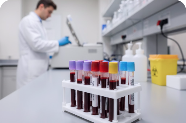
Analysis and Results
The extracted material is immediately prepared by smearing it onto glass slides and is often placed into a fixative solution. These slides are then sent to a cytology lab. There, a cytotechnologist screens the cells, and a specialist pathologist examines them in detail under a microscope. The study of these cells is called cytology.
The results are typically available within a few days and are categorized to indicate the findings:
Benign: No evidence of abnormal or cancerous cells.
Inflammatory: Evidence of infection or non-specific inflammation.
Atypical/Indeterminate: A mix of abnormal and normal cells, where the diagnosis is uncertain. This often necessitates follow-up testing, such as a core biopsy or surgical excision, to confirm the diagnosis.
Precancerous: Cells showing changes that could lead to cancer.
Malignant: Clear evidence of cancerous cells.
Based on the cytology results, the physician will explain the findings and discuss appropriate next steps, which may include further monitoring, targeted drug therapy, or surgery.
Safety, Risks, and Limitations
FNAC is considered a very safe procedure with infrequent complications. The risks are typically minor:
Minor Discomfort: Bruising, tenderness, or mild pain at the puncture site are the most common occurrences.
Infection or Bleeding: These are rare but possible risks.
Insufficient Sample: Occasionally, the sample obtained may not contain enough diagnostic material, requiring the test to be repeated. Inadequate rates are significantly lower when the samples are taken by expert cytopathologists or guided by imaging.
While FNAC is highly accurate when used appropriately, its main limitation compared to a core biopsy is that it only collects cells and fluid, not a larger core of tissue structure. This means FNAC is excellent for classifying cell types (cytology) but may sometimes provide less information on the architectural structure of the tissue, which can be essential for certain tumor gradings. For this reason, sometimes an FNAC result will prompt a follow-up core biopsy for confirmation or more detailed staging information.
Conclusion
FNAC is an indispensable diagnostic technique, offering a relatively quick, accurate, and minimally invasive way to investigate suspicious lumps. It allows for prompt diagnosis, helping clinicians and patients make timely decisions regarding treatment and ongoing management, thereby serving as a powerful initial step in the diagnostic pathway for a wide range of conditions.
Frequently Asked Questions (FAQs)
1. Is the FNAC test for TB?
Yes, the fine needle aspiration cytology test is a cost-effective, fast, and secure approach to diagnosing tuberculosis.
2. What happens if the FNAC test is positive?
A positive FNAC test result does not necessarily indicate a diagnosis of cancer. The physician may recommend additional tests to determine the definitive diagnosis, taking into account the patient's pre-existing medical condition, symptoms, complaints, and clinical examination.
3. What happens if the FNAC test is negative?
A negative result in an FNAC test report does not rule out the presence of the disease. An open biopsy should be conducted to confirm the diagnosis through histopathological examination.
4. What are some possible complications of the FNAC test?
Complications such as bleeding, infection, or bruising at the needle site may occur but are rare.
5. How long does the FNAC test take to perform?
While the test itself typically only takes a few minutes to complete, the entire appointment can last up to one hour, depending on the location of the mass and whether additional imaging tests are required.