


Understanding An Electroencephalogram
An Electroencephalogram, commonly known as an EEG, is a non-invasive diagnostic test used extensively in neurology to measure and record the electrical activity of the brain. Often described as a safe and painless procedure, the EEG provides healthcare professionals with critical insights into how the brain is functioning by tracking the electrical impulses used by brain cells (neurons) to communicate. This activity appears as distinctive wavy lines on the EEG recording, which change depending on whether a person is awake, asleep, or if abnormal brain activity is present.
What is an EEG?
The brain is constantly active, generating electrical signals even during sleep. These signals, or brain waves, are picked up by small, flat metal discs called electrodes, which are temporarily placed on the scalp. These electrodes are connected by wires to an EEG machine, which amplifies the delicate electrical signals and records them onto computer equipment or paper. The resulting record of brain activity is what healthcare providers analyze to look for characteristic patterns indicative of neurological conditions.
The technology behind the EEG has been utilized for many years and remains a fundamental tool in the diagnosis and management of a wide range of brain-related disorders. It offers a unique window into the brain's real-time electrical function, distinguishing it from imaging tests like MRI or CT scans which focus on brain structure.
Why is an EEG Performed?
The primary reason a healthcare provider orders an EEG is to detect abnormalities in brain activity. It is considered one of the main tests for diagnosing epilepsy and other seizure conditions, as seizure activity is characterized by distinctive, often sudden, changes in the brain’s electrical patterns.
Beyond seizure disorders, an EEG can play a vital role in diagnosing or monitoring various other neurological conditions, including:
Epilepsy and Seizure Disorders: Identifying the type of seizure and where it originates in the brain.
Brain Tumors: Detecting changes in brain activity around a growth.
Brain Damage: Assessing the extent of damage from a head injury (Traumatic Brain Injury).
Brain Infections: Such as encephalitis, where inflammation affects brain activity.
Sleep Disorders: Especially when conducted alongside a polysomnogram (Sleep EEG), to understand brain activity during sleep cycles.
Dementia/Alzheimer’s Disease: Evaluating the overall electrical state of the brain.
Coma: Used to monitor brain function in critically ill patients.
Confirming Brain Death: In specific clinical contexts, an EEG may be used to confirm the absence of electrical activity in the brain.
In some surgical settings, a continuous EEG may be used to monitor blood flow or to help find the right level of anesthesia for a patient in a medically induced coma.
Types of EEG Tests
Depending on the patient's condition and the information the provider needs, several types of EEG tests may be performed:
Routine EEG: Typically lasts between 20 and 40 minutes. During this standard test, the technician may ask the patient to perform simple actions like opening and closing their eyes, taking deep breaths (hyperventilation), or looking at flashing lights (photic stimulation), as these stimuli can sometimes trigger or highlight abnormal brain patterns.
Prolonged EEG: This test records brain activity for an hour or longer, increasing the chance of capturing intermittent or subtle electrical changes.
Sleep EEG: Brain wave patterns change significantly during sleep. For certain conditions, especially when seeking to diagnose a sleep-related seizure disorder, the test is specifically conducted while the patient is asleep. Patients may be asked to limit their sleep the night before to facilitate sleeping during the test.
Ambulatory EEG: This allows the patient to go about their normal daily routine while a small, portable recorder tracks brain activity over one or more days. This provides a long-term look at brain function in a natural environment.
Video EEG Monitoring (EEG Telemetry): In this specialized test, video and audio are recorded simultaneously with the EEG tracing. This helps providers correlate the patient's physical movements (like during a seizure) with the exact corresponding electrical activity in the brain, offering highly valuable diagnostic information. This is often done in an inpatient setting for several days.
The EEG Procedure: What to Expect
Preparing for an EEG is relatively simple, but following specific instructions is crucial for accurate results. Patients are usually asked to wash their hair the night before or the day of the test, avoiding the use of conditioners, hair sprays, gels, or creams, as these products can interfere with the adhesive used to attach the electrodes. Patients should also inform their provider of any medications they are taking.
On the day of the test:
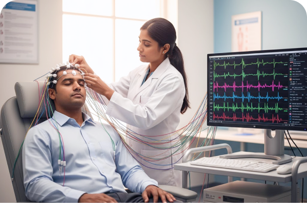
Preparation: A trained technician will measure the patient's head and mark specific points on the scalp where the electrodes will be placed.
Electrode Placement: Between 16 and 25 small electrodes are attached to the scalp using a special glue, paste, or adhesive. In some cases, a fitted elastic cap with embedded electrodes may be used.
The Recording: The patient will be asked to sit in a comfortable reclining chair or lie on a bed, relax, and remain still with their eyes closed or open, as directed. The electrodes send the brain's electrical signals to the EEG machine, which records the wave patterns.
Stimulation (Routine EEG): The technician may administer simple stimuli, such as asking the patient to breathe deeply for a few minutes or exposing them to flashing lights, to check for specific responses in the brain activity.
A routine EEG usually takes less than an hour, typically between 20 and 40 minutes, once the electrodes are in place. Longer tests are scheduled for sleep studies or prolonged monitoring.
Safety and Results
An EEG is considered a safe and painless procedure. The electrodes simply record activity and do not transmit any electrical current to the body.
In rare instances, for individuals with epilepsy, the hyperventilation or flashing lights used during the test may intentionally trigger a seizure. However, this is done in a controlled clinical environment with medical staff present to provide immediate care.
Following the test, a neurologist or healthcare professional trained in EEG interpretation will analyze the recorded brain wave patterns. They look for abnormal patterns, spikes, or slowdowns in activity that could indicate a neurological problem. The results are then used to guide diagnosis, inform treatment plans, and monitor the effectiveness of existing therapies