


Understanding A Computerized Tomography (CT) scan
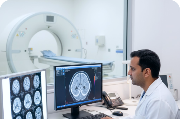
A Computerized Tomography (CT) scan of the brain, sometimes called a brain CAT scan, is a noninvasive imaging procedure that uses X-rays and a computer to create detailed cross-sectional images ("slices") of the head. These images provide clear views of the brain, bone, blood vessels, and soft tissues.
What is a Brain CT Scan Used to Detect?
The primary use of a brain CT scan is for quick imaging, making it the initial test after head injuries or sudden signs of a stroke. It provides rapid and critical information about the brain's condition.
A brain CT scan can be used to detect and evaluate:
Emergencies: Head trauma, bleeding in the brain, stroke (including blood clots), and fluid buildup (hydrocephalus).
Structural Issues: Skull fractures, structural brain issues, and sinus-related bone issues.
Diseases and Conditions: Tumors, infections, brain diseases, and the effects of treatment on brain tumors.
Symptoms: Causes of sudden symptoms such as severe headaches, loss of consciousness, seizures, and weakness.
Types of Brain CT Scans
Depending on the medical need, a CT scan may be performed with or without the use of a contrast agent:
Noncontrast CT Scan: This is the fastest type of CT imaging and is typically used in emergencies to evaluate conditions like head trauma or bleeding, as these conditions are often better seen without contrast.
Contrast-Enhanced CT Scan: A contrast agent (dye) is injected into a vein, which helps highlight specific areas, such as blood vessels or certain types of tumors, for further evaluation.
CT Angiography (CTA): This specialized test uses contrast to focus on the blood vessels, helping to find aneurysms (bulging blood vessels), arteriovenous malformations, or blood clots.
CT Perfusion Scan: Measures blood flow in the brain, which can help show brain tissue that might be saved after a stroke (ischemic penumbra).
Preparation for the Procedure
Preparation steps ensure the best possible image quality and patient safety:
Fasting: You may be asked not to eat or drink for a few hours before your scheduled exam to avoid potential complications like nausea. Medications can generally be taken with a small sip of water, but follow your doctor’s specific instructions.
Clothing and Metal Objects: You may be asked to change into a hospital gown. All metal objects—including belts, jewelry, dentures, and eyeglasses—must be removed as they can interfere with the X-ray images.
Contrast Media: If a contrast-enhanced scan is required, you will be asked about any allergies or previous reactions to contrast agents, as well as any kidney conditions. You will sign a consent form detailing the associated risks and side effects. Inform your healthcare professional if you are pregnant or breastfeeding.
What Happens During a Brain CT Scan?
The procedure is quick and generally takes about 15 to 30 minutes, though preparation time will vary.
Positioning: You will lie on a movable scan table that slides into the large, circular opening of the CT scanning machine.
Contrast Injection (if needed): If contrast is being used, an IV line will be started in your hand or arm for the injection.
Scanning: The machine rotates around your head, taking X-ray images from multiple angles. You must remain very still during the procedure to prevent blurry images. You will hear clicking sounds as the scanner operates.
Image Creation: A computer receives the X-ray data and transforms the information into cross-sectional images for interpretation by a radiologist.
How does a CT scan work?
Healthcare providers use CT scans to see things that regular X-rays can’t show. It produces detailed, clear, and precise images of the organs and structures in your body. To get these images, a CT machine takes X-ray pictures as it moves around you.
X-rays alone take flat, 2D images. A CT scan takes several pictures at many angles to create cross-sectional images. Just like you can see the inside layers of a cake when you slice it, a CT can show the “layers” of your body. Taken together, the layers create a 3D image. Some CT scans use a contrast material (dye) to make the pictures even clearer.
What should I expect during my CT scan?
During the test, you’ll usually lie on your back on a table (like a bed). When the scan begins:
The bed moves into the doughnut-shaped machine. At this point, you’ll need to stay as still as possible because movement can blur the images.
You may also be asked to hold your breath for a short period of time, usually fewer than 15 to 20 seconds.
The scanner takes pictures of the area your healthcare provider needs to see. A CT scanner is relatively quiet.
When the exam is over, the table moves back out of the scanner.
A technologist trained specifically to perform CT scans will be there to guide you through the entire process.
Are there risks or side effects?
Healthcare providers consider CT scans safe. CT scans for children are safe, too. Like X-rays, CT scans use a small amount of ionizing radiation to capture images. The level of radiation you’re exposed to is small. If you have concerns about the health risks of CT scans, talk to your healthcare provider. They’ll help you make an informed decision about the scan.
CT scans themselves usually don’t cause side effects. But some people have side effects from the contrast material. These side effects may include:
What do the results of a CT scan mean?
It depends on what your healthcare provider was looking for. The report your radiologist writes may include things like:
How they performed the CT (if contrast was used, positioning, what body parts they looked at)
Any findings (descriptions of any irregularities or details as to what looked normal or abnormal)
A summary called an Impression (including a possible diagnosis, the biggest finding, and recommendations for further study)
Sometimes, your provider will suggest additional testing after a CT. For example, they may recommend an MRI to get a different look at a suspicious area.
Safety and Risks
Brain CT scans are generally considered safe. They use a low, carefully controlled amount of radiation, and the benefits of finding a serious brain problem typically outweigh the minimal potential risks. For children, lower doses of radiation are used, and scans are performed only when medically necessary due to their increased sensitivity.
If contrast is used, there is a small risk of allergic reaction. Patients with certain conditions, such as kidney disease, may face greater risks with contrast media, which is why a blood test to check kidney function may be required beforehand.
Frequently Asked Questions (FAQs)
1. Can a CT scan detect cancer?
Yes. It helps detect cancer because it shows tumors, abnormal growths or changes in your organs that could be cancer. Providers also use CT to see if cancer has spread or if treatment is working.
2. Can I have a CT scan if I’m pregnant?
If you’re pregnant or think you might be pregnant, you should tell your provider. CT scans of your pelvis and abdomen can expose the fetus to radiation, but it’s not enough to cause harm. CT scans in other parts of your body have almost no radiation to the fetus.