


2D Echocardiography (2D Echo): A Detailed Look at Your Heart
A 2D echocardiography, commonly referred to as a 2D echo, is a non-invasive, painless ultrasound scan of the heart. It uses high-frequency sound waves to create moving, two-dimensional images of the heart’s chambers, valves, and nearby blood vessels. This test helps doctors see how well your heart is both structured and functioning in real time.
What is a 2D Echocardiography?
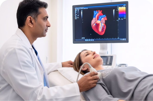
A 2D echo works like an ultrasound used in pregnancy, but it is focused on the heart. A device called a transducer sends sound waves into the chest. These waves bounce off the heart structures and return to the transducer. A computer then converts the returning signals into live images on a monitor.
The 2D images usually appear as cone-shaped cross-sections, showing the motion of the heart walls and valves. When combined with Doppler techniques, the test can also show how blood is flowing through the heart and valves.
Why is a 2D Echo Performed?
Doctors may recommend a 2D echo to:
Check how well the heart pumps blood, including measuring the ejection fraction of the left ventricle.
Evaluate heart valves for leakage (regurgitation) or narrowing (stenosis).
Detect structural problems such as thickened heart muscle, congenital heart defects, or fluid buildup around the heart (pericardial effusion).
Investigate symptoms like chest pain, shortness of breath, palpitations, fainting, or heart murmurs.
Monitor known heart conditions and track the effectiveness of treatments or surgery.
The Procedure: What to Expect
A 2D echo is usually done in a hospital, clinic, or cardiology lab and takes about 30 to 60 minutes.
Before the test:
In most cases, no special preparation is needed. You can eat and take your medications as usual, unless your doctor gives specific instructions. You will be asked to undress from the waist up and wear a gown.During the test:
You lie on an examination table, usually on your back or left side.
Electrodes are placed on your chest to record your heart’s electrical activity and synchronise it with the images.
A clear gel is applied to your chest to help the sound waves travel.
The sonographer presses the transducer on different parts of your chest and moves it around to capture images from various angles. You may feel mild pressure and may be asked to change position or briefly hold your breath.
If Doppler is used, you may hear a whooshing sound, which represents blood flow through the heart.
After the test:
The gel is wiped off, and you can return to your normal activities immediately. A cardiologist reviews the images and prepares a report for your doctor.
2D Echo vs. 3D Echo
While 3D echocardiography can provide more detailed three-dimensional views for specific complex problems, the 2D echo remains the standard, widely used test for routine assessment of heart structure and function. It offers excellent information for most clinical needs.
Overall, 2D echocardiography is a safe, informative, and essential tool in cardiology, helping ensure accurate diagnosis and effective management of heart conditions.
Frequently Asked Questions (FAQs)
1. Do I need to fast before a 2D echo?
For a standard 2D echocardiogram, fasting is not required. You can eat, drink, and take your medications normally.
2. How long does it take to get results?
Results are typically delivered within 24-48 hours.
3. Are there any risks or side effects?
The standard transthoracic 2D echo has no known risks and is considered extremely safe. There's no radiation exposure, and the ultrasound waves are harmless. You can return to normal activities immediately after the test.